07 · Cartilage Repair
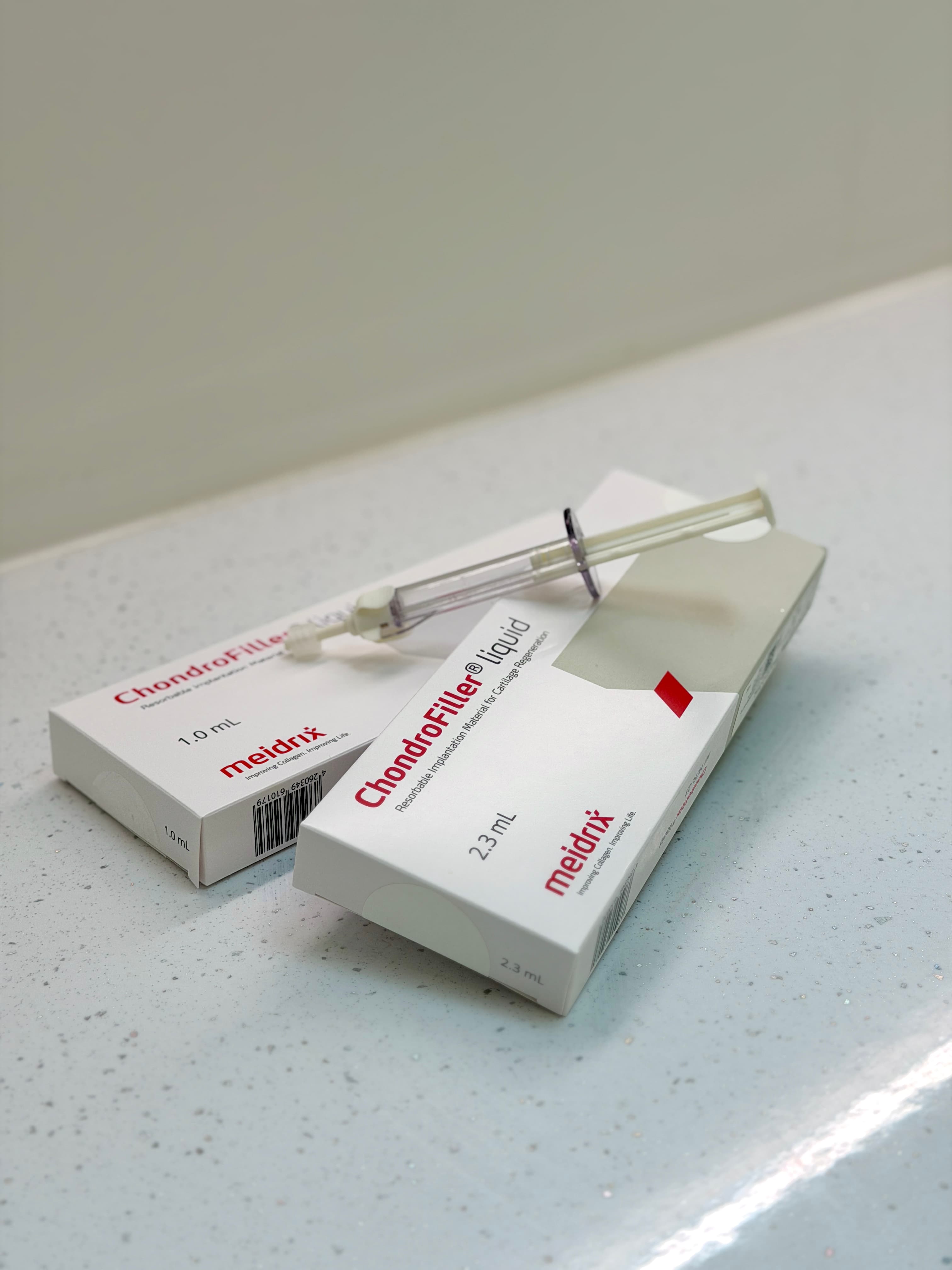
ChondroFiller for Knee Cartilage Defects
Early Clinical Outcomes of a Single-Stage Collagen Gel
A retrospective review of 42 knees treated arthroscopically with a cell-free collagen gel in one sitting. KOOS rose from 58.1 to 84.5 and activity pain fell from 7 to 2.3, with 79% reaching the minimal clinically important difference.
Presentation Details
Presented, and open to scrutiny.
Each presentation connects surgical decision-making, markerless movement measurement and quantitative imaging — bringing MSK Doctors Research to an international clinical and scientific audience.
Event
BOA Annual Congress 2026, Olympia London
Format
A0 e-poster
Authors
Tanvi Verma · Paul Lee — MSK Doctors, Sleaford, UK and London Cartilage Clinic, London, UK; Selin Demirel · Elif İrem Kılıç · Ahmet Burak Çelik · Feza Korkusuz — Hacettepe University, Sports Medicine, Ankara, Türkiye
Themes
Scientific Focus
Articular cartilage has limited intrinsic repair capacity, so untreated focal defects may enlarge and contribute to premature osteoarthritis and eventual arthroplasty. Established restorative techniques can require cell harvest, staged procedures, graft fixation or prolonged rehabilitation. ChondroFiller is an absorbable, cell-free type I collagen gel delivered arthroscopically after defect preparation: it conforms to the lesion and hardens in situ, providing a three-dimensional scaffold intended to support endogenous repair in a single procedure. This single-centre retrospective review covered 40 patients (42 knees) with MRI-confirmed ICRS grade III–IV defects and at least 12 months of follow-up, excluding diffuse osteoarthritis, major malalignment and prior restoration at the index site.
Why It Matters
KOOS improved from 58.1 to 84.5 and activity VAS pain fell from 7 to 2.3, both at p < 0.001. Seventy-nine per cent achieved the minimal clinically important difference and 64% returned to their preferred sport or recreation. Follow-up MRI at a mean of 14.6 months showed complete or nearly complete defect fill in 70% of cases. The proportions are descriptive rather than inferential, and follow-up dispersion was not reported in the source deck — the authors state that larger comparative studies are required to assess durability and optimal indications.
Next in the programme
ChondroFiller for Ankle Cartilage Defects
Clinical and Imaging Outcomes in Focal Talar Dome Lesions