08 · Cartilage Repair
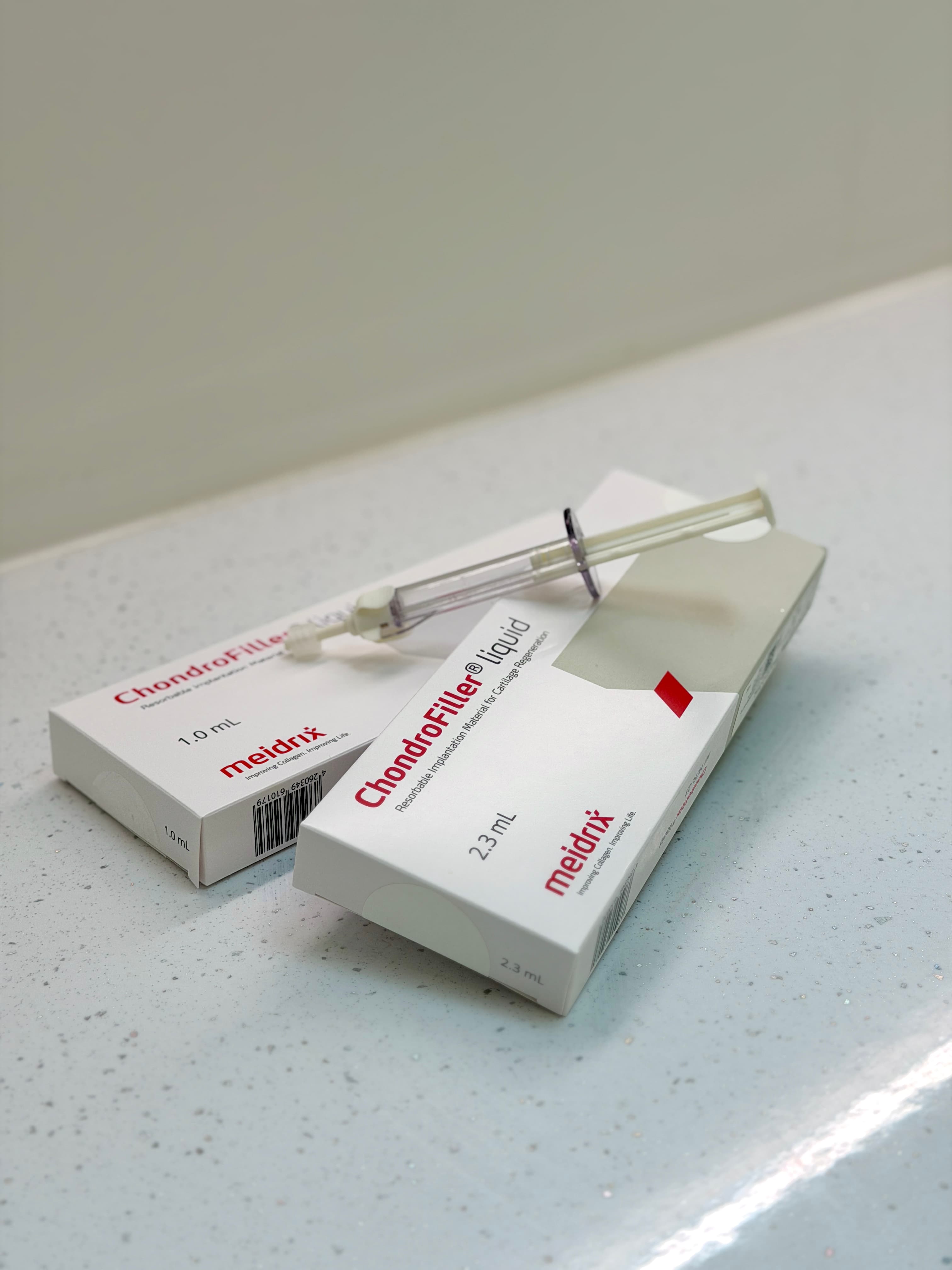
ChondroFiller for Ankle Cartilage Defects
Clinical and Imaging Outcomes in Focal Talar Dome Lesions
Thirty ankles treated arthroscopically with a cell-free collagen gel. FAOS rose from 59.8 to 86.7 and activity pain fell from 6.7 to 2.1, with complete or nearly complete defect fill on 68% of follow-up MRIs.
Presentation Details
Presented, and open to scrutiny.
Each presentation connects surgical decision-making, markerless movement measurement and quantitative imaging — bringing MSK Doctors Research to an international clinical and scientific audience.
Event
BOA Annual Congress 2026, Olympia London
Format
A0 e-poster
Authors
Tanvi Verma · Paul Lee — MSK Doctors, Sleaford, UK; London Cartilage Clinic, London, UK; Selin Demirel · Elif İrem Kılıç · Ahmet Burak Çelik · Feza Korkusuz — Hacettepe University, Sports Medicine, Ankara, Türkiye
Themes
Scientific Focus
Cartilage defects commonly follow ankle sprain, fracture or repetitive microtrauma. Because articular cartilage heals poorly, these lesions can cause persistent pain, swelling and functional limitation, and may progress to early post-traumatic osteoarthritis. Existing restorative procedures may require graft harvest, staged treatment or more invasive surgery. This single-centre retrospective cohort covered 30 ankles in 29 adults treated arthroscopically over 24 months, requiring an MRI-confirmed focal talar dome defect, persistent symptoms despite at least three months of non-operative care, and at least 12 months of follow-up. The defect was debrided to stable margins before the collagen gel filled it and hardened in situ.
Why It Matters
FAOS improved from 59.8 to 86.7 and activity VAS pain fell from 6.7 to 2.1 (p < 0.001). Seventy-three per cent of patients were satisfied and 61% returned to activity, while follow-up MRI at a mean of 13.5 months showed complete or nearly complete defect fill in 68% of the 22 ankles imaged. No deep infections, graft reactions or thromboembolic events were reported. The cohort is retrospective, single-centre and without a comparator, and MRI was available for 22 of the 30 ankles — larger comparative studies with longer follow-up are required to define durability and optimal indications.
Next in the programme
Knee OA Severity and Hip Movement Quality
C.R.A.F.T. Scores Across Kellgren–Lawrence Grades in 177 Patients